|
|
Adrenal Myelolipoma (Myolipoma)
General Considerations
- Rare, usually non-functioning tumors found incidentally most often in males aged 40-60
- Benign tumors composed of fat and hemopoietic cells enclosed by pseudocapsule
- Usually occurs in adrenal gland but may be extra-adrenal in 15%
Clinical Findings
- Almost always asymptomatic
- Most are discovered incidentally during a CT scan for other reasons
- Adrenal masses are discovered incidentally in about 2.5-5% of abdominal CT scans
- May be painful if there is hemorrhage
Imaging Findings
- Unilateral
- Usually small (<4 cm)
- May be large in size (~10 com)
- Hemorrhage is more common in larger lesions
- Calcifications can occur in ¼ from prior hemorrhage
- CT
- Fatty mass of -30 to -115 HU
- Fat interspersed with higher attenuation marrow-like elements
- US
- Mostly hyperechoic mass with interspersed hypoechoic regions
- MRI
- Heterogeneously high signal on T1
- Intermediate intensity similar to spleen on T2
- In opposed phase, there is a drop in signal intensity
Differential Diagnosis
- Adrenal cysts
- Water density with thin wall
- Adrenal adenomas
- Most are fat containing with densities of <10 HU
- Density measurements before and after contrast administration can be used
- Enhancement washout (HU portal venous phase minus HU delayed phase)/ HU portal venous phase x 100
- If value is > 50%, adrenal adenoma
- Sensitivity of 93% and specificity 98%
Treatment
- Conservative with follow-up to determine if there is enlargement for masses >4 cm
- Symptomatic masses are usually surgically removed
Complications
- Occasionally associated with endocrine disturbances
- Conn’s syndrome
- Addison’s disease
- Acute hemorrhage may be associated with increase in size

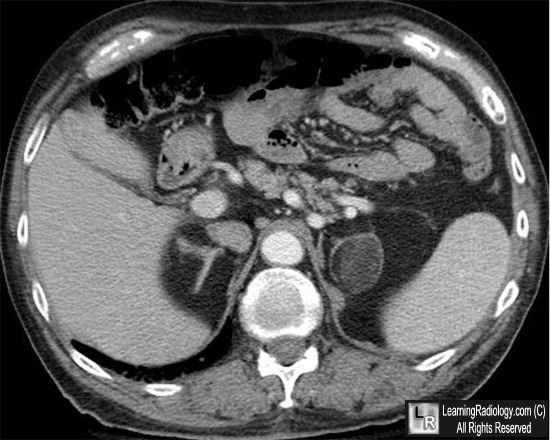
Adrenal myelipoma. Contrast-enhanced axial CT scan image through the upper abdomen reveals a large, partially fat-containing mass in the left adrenal glad (white arrow). The mass has a thin capsule surrounding it. A portion of the normal right adrenal is seen (red arrow).
For more information, click on the link if you see this icon 
For this same photo without the annotations, click here
Kohli A. Adrenal Incidentilomas: Can we characterize them?. Indian J Radiol Imaging 2006;16:163-4
|
|
|
